Under the Surface, the Brain Seethes With Undiscovered Activity
University of Rochester researchers have found that roughly 80 percent of our cognitive power may be cranking away on tasks completely unknown to us, probably dedicated to subconsciously reprocessing our initial thoughts and experiences. The research has possible profound implications for our very basis of understanding reality.
There’s an old myth that we only use 10 percent of our brains, but researchers at the University of Rochester have found in reality that roughly 80 percent of our cognitive power may be cranking away on tasks completely unknown to us. Curiously, this clandestine activity does not exist in the youngest brains, leading scientists to believe that the mysterious goings-on that absorb the majority of our minds are dedicated to subconsciously reprocessing our initial thoughts and experiences. The research, which has possible profound implications for our very basis of understanding reality, appears in this week’s issue of the journal Nature.
“We found neural activity that frankly surprised us,” says Michael Weliky, associate professor of brain and cognitive sciences at the University of Rochester. “Adult ferrets had neural patterns in their visual cortex that correlated very well with images they viewed, but that correlation didn’t exist at all in very young ferrets, suggesting the very basis of comprehending vision may be a very different task for young brains versus old brains.”
A second surprise was in store for Weliky. Placing the ferrets in a darkened room revealed that older ferrets’ brains were still humming along at 80 percent as if they were processing visual information. Since this activity was absent in the youngsters, Weliky and his colleagues were left to wonder: What is the visual cortex so busy processing when there’s no image to process?
Initially, Weliky’s research was aimed at studying whether visual processing bore any resemblance to the way real-world images appear. This finding may help lead to a better understanding of how neurons decode our world and how our perception of reality is shaped.
Weliky, in a bit of irony, set 12 ferrets watching the reality-stretching film The Matrix. He recorded how their brains responded to the film, as well as to a null pattern like enlarged television static, and a darkened room. Movies capture the visual elements that are present in the real world. For instance, as Keanu’s hand moves across the screen for a karate chop, the image of the hand and all the lines and color it represents moves across a viewer’s visual realm essentially the same way it would in real life. By contrast, the enlarged static—blocks of random black and white—has no such motion. Weliky was able to graph the movie-motion statistically, showing essentially how objects move in the visual field.
The test was then to see if there was any relationship between the statistical motion of the movie and the way visual neurons in the ferrets fired. Each visual neuron is keyed to respond to certain visual elements, such as a vertical line, that appears in a specific area of the ferret’s vision. A great number of these cells combine to process an image of many lines, colors, etc. By watching the patterns of how these cells fired while watching The Matrix, Weliky could describe the pattern statistically, and match those statistics of how the ferret responded to the film with the statistics of the actual visual aspects of the film.
Weliky found two surprises. First, while the neurons of adult ferrets statistically seemed to respond similarly to the statistics of the film itself, younger ferrets had almost no relationship. This suggests that though the young ferrets are taking in and processing visual stimuli, they’re not processing the stimuli in a way that reflects reality.
“You might think of this as a sort of dyslexia,” explains Weliky. “It may be that in very young brains, the processing takes place in a way that’s not necessarily disordered, but not analogous to how we understand reality to be. It’s thought that dyslexia works somewhat like this—that some parts of the brain process written words in an unusual way and seem to make beginnings of words appear at their ends and vice versa. Infant brains may see the entire world the same way, as a mass of disparate scenes and sounds.” Weliky is quick to point out that whatever way infant brains may interpret the world, just because they’re different from an adult pattern of perception does not mean the infants have the wrong perception. After all, an adult interpreted the visual aspects of the film with our adult brains, so it shouldn’t be such a surprise that other adult brains simply interpret the visual aspects the same way. If an infant drew up the statistics, it might very well match the neural patterns of other infants.
The second, and more surprising, result of the study came directly from the fact that Weliky’s research is one of the first to test these visual neurons while the subject is awake and watching something. In the past, researchers would perhaps shine a light at an unconscious ferret and note which areas of the brain responded, but while that method narrowed the focus to how a single cell responds, it eliminated the chance to understand how the neural network of a conscious animal would respond. Accepting all the neural traffic of a conscious brain as part of the equation let Weliky get a better idea of the actual processing going on. As it turned out, one of his control tests yielded insight into neural activity no one expected.
When the ferrets were in a darkened room, Weliky expected their visual neurons to lack any kind of activity that correlated with visual reality. Neurologists have long known that there is substantial activity in the brain, even in darkness, but the pattern of that activity had never been investigated. Weliky discovered that while young ferrets displayed almost no patterns that correlated with visual reality, the adult ferrets’ brains were humming along, producing the patterns even though there was nothing to see. When watching the film, the adult ferrets’ neurons increased their patterned activity by about 20 percent.
“This means that in adults, there is a tremendous amount of real-world processing going on—80 percent—when there is nothing to process,” says Weliky. “We think that if you’ve got your eyes closed, your visual processing is pretty much at zero, and that when you open them, you’re running at 100 percent. This suggests that with your eyes closed, your visual processing is already running at 80 percent, and that opening your eyes only adds the last 20 percent. The big question here is what is the brain doing when it’s idling, because it’s obviously doing something important.”
Since the young ferrets do not display similar patterns, the “idling” isn’t necessary for life or consciousness, but since it’s present in the adults even without stimulus, Weliky suggests it may be in a sense what gives the ferret its understanding of reality. The eye takes in an image and the brain processes the image, but 80 percent of the activity may be a representation of the world replicated inside the ferret’s brain.
“The basic findings are exciting enough, but you can’t help but speculate on what they might mean in a deeper context,” says Weliky. “It’s one thing to say a ferret’s understanding of reality is being reproduced inside his brain, but there’s nothing to say that our understanding of the world is accurate. In a way, our neural structure imposes a certain structure on the outside world, and all we know is that at least one other mammalian brain seems to impose the same structure. Either that or The Matrix freaked out the ferrets the way it did everyone else.”
This research was funded by the National Institutes of Health.

04 October 2004
Testing Bell's inequality in a capacitively coupled Josephson circuit L.F. Wei, Yu-xi Liu, Franco Nori quant-ph 0408089 "Bell's inequalities have been experimentally tested by using, e.g., far apart photons and very-closely-spaced trapped ions. Here, we propose a way to test Bell's inequality with a pair of capacitively Josephson qubits; these coupled-qubits exhibit macroscopic quantum entanglement as demonstrated by recent spectral-analysis experiments [Nature 421, 823 (2003); Science 300, 1548 (2003)]. We propose an effective dynamical decoupling approach to overcome the "fixed-interaction" difficulty for implementing the required single-qubit operations. The obtained long-lived entanglement and realizable simultaneous measurements of the two qubits should allow the testing of Bell's inequality using this coupled Josephson circuit."
21 September 2004
Quantum entanglement by classical computer: a crucial experiment
Luigi Accardi, Centro V. Volterra, Roma
A simple experiment is described in which two experimenters, by performing independent, local, binary choices on a common classical, deterministic, macroscopic source of randomness (in fact a generator of random points in the unit disk in the plane) and computing the empirical correlations among their results, arrive to a violation of Bell's inequalities. The local binary choices satisfy all the standard conditions of the EPR experiment: singlet, equiprobability, rotation invariance, etc.
In addition the experiment suggests a new interpretation of the usual EPR experiment, more natural and appealing from the physical point of view than the usual one and totally in line with the "chameleon effect" which is at the basis of the quantum probabilistic approach to the theory of quantum measurement.
A mathematical formulation of the "chameleon effect" will be discussed and illustrated with the mathematical model used to write the computer programme used in the experiment. The result of the present experiment, which for a long time has been considered to be impossible by the majority of physicists, fully confirms the point of view advocated, starting from the late 70's, by quantum probability in absolute isolation and strongly opposed by the majority of physicists who, following the interpretation due to Bell, were relating the violation of Bell's inequality to a non locality effect.
In particular the experiment proves that:
(i) it is possible to produce non-Kolmogorovian correlations by local realistic classical deterministic macroscopic systems
(ii) it is possible to produce quantum entanglement by classical
computer
This opens the way to a series of new possibilities, for example the possibility of implementing quantum cryptography by classical computer. The experiment will be described and performed during the talk and the public will have the possibility to check the procedure by choosing the parameters of the measurements. An earlier version of the experiment is available at the Volterra Institute.
Luigi Accardi, Centro V. Volterra, Roma
A simple experiment is described in which two experimenters, by performing independent, local, binary choices on a common classical, deterministic, macroscopic source of randomness (in fact a generator of random points in the unit disk in the plane) and computing the empirical correlations among their results, arrive to a violation of Bell's inequalities. The local binary choices satisfy all the standard conditions of the EPR experiment: singlet, equiprobability, rotation invariance, etc.
In addition the experiment suggests a new interpretation of the usual EPR experiment, more natural and appealing from the physical point of view than the usual one and totally in line with the "chameleon effect" which is at the basis of the quantum probabilistic approach to the theory of quantum measurement.
A mathematical formulation of the "chameleon effect" will be discussed and illustrated with the mathematical model used to write the computer programme used in the experiment. The result of the present experiment, which for a long time has been considered to be impossible by the majority of physicists, fully confirms the point of view advocated, starting from the late 70's, by quantum probability in absolute isolation and strongly opposed by the majority of physicists who, following the interpretation due to Bell, were relating the violation of Bell's inequality to a non locality effect.
In particular the experiment proves that:
(i) it is possible to produce non-Kolmogorovian correlations by local realistic classical deterministic macroscopic systems
(ii) it is possible to produce quantum entanglement by classical
computer
This opens the way to a series of new possibilities, for example the possibility of implementing quantum cryptography by classical computer. The experiment will be described and performed during the talk and the public will have the possibility to check the procedure by choosing the parameters of the measurements. An earlier version of the experiment is available at the Volterra Institute.
15 September 2004
Chips Coming to a Brain Near You
In this era of high-tech memory management, next in line to get that memory upgrade isn't your computer, it's you.
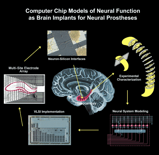
Professor Theodore W. Berger , director of the Center for Neural Engineering at the University of Southern California, is creating a silicon chip implant that mimics the hippocampus, an area of the brain known for creating memories. If successful, the artificial brain prosthesis could replace its biological counterpart, enabling people who suffer from memory disorders to regain the ability to store new memories.
And it's no longer a question of "if" but "when." The six teams involved in the multi-laboratory effort, including USC, the University of Kentucky and Wake Forest University, have been working together on different components of the neural prosthetic for nearly a decade. They will present the results of their efforts at the Society for Neuroscience 's annual meeting in San Diego, which begins Saturday.
While they haven't tested the microchip in live rats yet, their research using slices of rat brain indicates the chip functions with 95 percent accuracy. It's a result that's got the scientific community excited.
"It's a new direction in neural prosthesis," said Howard Eichenbaum , director of the Laboratory of Cognitive Neurobiology at Boston University. "The Berger enterprise is ambitious, aiming to provide a prosthesis for memory. The need is high, because of the prevalence of memory disorder in aging and disease associated with loss of function in the hippocampus."
Forming new long-term memories may involve such tasks as learning to recognize a new face, or remembering a telephone number or directions to a new location. Success depend on the proper functioning of the hippocampus. While this part of the brain doesn't store long-term memories, it re-encodes short-term memory so it can be stored as long-term memory.
It's the area that's often damaged as a result of head trauma, stroke, epilepsy and neurodegenerative disorders such as Alzheimer's disease. Currently, no clinically recognized treatments exist for a damaged hippocampus and the accompanying memory disorders.
Berger's team began its research by studying the re-encoding process performed by neurons in slices of rat hippocampi kept alive in nutrients. By stimulating these neurons with randomly generated computer signals and studying the output patterns, the group determined a set of mathematical functions that transformed any given arbitrary input pattern in the same manner that the biological neurons do. And according to the researchers, that's the key to the whole issue.
"It's an impossible task to figure out what your grandmother looks like and how I would encode that," said Berger. "We all do a lot of different things, so we can't create a table of all the things we can possibly look at and how it's encoded in the hippocampus. What we can do is ask, 'What kind of transformation does the hippocampus perform?'
"If you can figure out how the inputs are transformed, then you do have a prosthesis. Then I could put that into somebody's brain to replace it, and I don't care what they look at -- I've replaced the damaged hippocampus with the electronic one, and it's going to transform inputs into outputs just like the cells of the biological hippocampus."
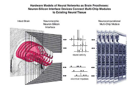
Dr. John J. Granacki , director of the Advanced Systems Division at USC, has been working on translating these mathematical functions onto a microchip. The resulting chip is meant to simulate the processing of biological neurons in the slice of rat hippocampus: accepting electrical impulses, processing them and then sending on the transformed signals. The researchers say the microchip is doing exactly that, with a stunning 95 percent accuracy rate.
"If you were looking at the output right now, you wouldn't be able to tell the difference between the biological hippocampus and the microchip hippocampus," Berger said. "It looks like it's working."
The team next plans to work with live rats that are moving around and learning, and will study monkeys later. The researchers will investigate drugs or other means that could temporarily deactivate the biological hippocampus, and implant the microchip on the animal's head, with electrodes into its brain.
"We will attempt to adapt the artificial hippocampus to the live animal and then show that the animal's performance -- dependent in these tasks on an intact hippocampus -- will not be compromised when the device is in place and we temporarily interrupt the normal function of the hippocampus," said Sam A. Deadwyler , "thus allowing the neuro-prosthetic device to take over that normal function." Deadwyler, a professor at Wake Forest University, is working on measuring the hippocampal neuron activity in live rats and monkeys.
The team expects it will take two to three years to develop the mathematical models for the hippocampus of a live, active rat and translate them onto a microchip, and seven or eight years for a monkey. They hope to apply this approach to clinical applications within 10 years. If everything goes well, they anticipate seeing an artificial human hippocampus, potentially usable for a variety of clinical disorders, in 15 years.
Overall, experts find the results promising.
"We are nowhere near applicability," said Boston University's Eichenbaum. "But the next decade will prove whether this strategy is truly feasible."
"There is a big gap in making the microchip work in a slice preparation and getting it to work in a human being," added Norbert Fortin, a neuroscientist from the Cognitive Neurobiology Lab at Boston University. "However, their approach is very methodical, and it is not unreasonable to think that in 15 to 20 years such a chip could help, to some degree, a patient who suffered from hippocampal damage."
Research Group, Wired Link
In this era of high-tech memory management, next in line to get that memory upgrade isn't your computer, it's you.
Professor Theodore W. Berger , director of the Center for Neural Engineering at the University of Southern California, is creating a silicon chip implant that mimics the hippocampus, an area of the brain known for creating memories. If successful, the artificial brain prosthesis could replace its biological counterpart, enabling people who suffer from memory disorders to regain the ability to store new memories.
And it's no longer a question of "if" but "when." The six teams involved in the multi-laboratory effort, including USC, the University of Kentucky and Wake Forest University, have been working together on different components of the neural prosthetic for nearly a decade. They will present the results of their efforts at the Society for Neuroscience 's annual meeting in San Diego, which begins Saturday.
While they haven't tested the microchip in live rats yet, their research using slices of rat brain indicates the chip functions with 95 percent accuracy. It's a result that's got the scientific community excited.
"It's a new direction in neural prosthesis," said Howard Eichenbaum , director of the Laboratory of Cognitive Neurobiology at Boston University. "The Berger enterprise is ambitious, aiming to provide a prosthesis for memory. The need is high, because of the prevalence of memory disorder in aging and disease associated with loss of function in the hippocampus."
Forming new long-term memories may involve such tasks as learning to recognize a new face, or remembering a telephone number or directions to a new location. Success depend on the proper functioning of the hippocampus. While this part of the brain doesn't store long-term memories, it re-encodes short-term memory so it can be stored as long-term memory.
It's the area that's often damaged as a result of head trauma, stroke, epilepsy and neurodegenerative disorders such as Alzheimer's disease. Currently, no clinically recognized treatments exist for a damaged hippocampus and the accompanying memory disorders.
Berger's team began its research by studying the re-encoding process performed by neurons in slices of rat hippocampi kept alive in nutrients. By stimulating these neurons with randomly generated computer signals and studying the output patterns, the group determined a set of mathematical functions that transformed any given arbitrary input pattern in the same manner that the biological neurons do. And according to the researchers, that's the key to the whole issue.
"It's an impossible task to figure out what your grandmother looks like and how I would encode that," said Berger. "We all do a lot of different things, so we can't create a table of all the things we can possibly look at and how it's encoded in the hippocampus. What we can do is ask, 'What kind of transformation does the hippocampus perform?'
"If you can figure out how the inputs are transformed, then you do have a prosthesis. Then I could put that into somebody's brain to replace it, and I don't care what they look at -- I've replaced the damaged hippocampus with the electronic one, and it's going to transform inputs into outputs just like the cells of the biological hippocampus."
Dr. John J. Granacki , director of the Advanced Systems Division at USC, has been working on translating these mathematical functions onto a microchip. The resulting chip is meant to simulate the processing of biological neurons in the slice of rat hippocampus: accepting electrical impulses, processing them and then sending on the transformed signals. The researchers say the microchip is doing exactly that, with a stunning 95 percent accuracy rate.
"If you were looking at the output right now, you wouldn't be able to tell the difference between the biological hippocampus and the microchip hippocampus," Berger said. "It looks like it's working."
The team next plans to work with live rats that are moving around and learning, and will study monkeys later. The researchers will investigate drugs or other means that could temporarily deactivate the biological hippocampus, and implant the microchip on the animal's head, with electrodes into its brain.
"We will attempt to adapt the artificial hippocampus to the live animal and then show that the animal's performance -- dependent in these tasks on an intact hippocampus -- will not be compromised when the device is in place and we temporarily interrupt the normal function of the hippocampus," said Sam A. Deadwyler , "thus allowing the neuro-prosthetic device to take over that normal function." Deadwyler, a professor at Wake Forest University, is working on measuring the hippocampal neuron activity in live rats and monkeys.
The team expects it will take two to three years to develop the mathematical models for the hippocampus of a live, active rat and translate them onto a microchip, and seven or eight years for a monkey. They hope to apply this approach to clinical applications within 10 years. If everything goes well, they anticipate seeing an artificial human hippocampus, potentially usable for a variety of clinical disorders, in 15 years.
Overall, experts find the results promising.
"We are nowhere near applicability," said Boston University's Eichenbaum. "But the next decade will prove whether this strategy is truly feasible."
"There is a big gap in making the microchip work in a slice preparation and getting it to work in a human being," added Norbert Fortin, a neuroscientist from the Cognitive Neurobiology Lab at Boston University. "However, their approach is very methodical, and it is not unreasonable to think that in 15 to 20 years such a chip could help, to some degree, a patient who suffered from hippocampal damage."
Research Group, Wired Link
Subscribe to:
Posts (Atom)